The precision of T1 hypointense lesion volume quantification in multiple sclerosis treatment trials: A multicenter study. 2007. Fartaria MJ, Bonnier G, Roche A, Kober T, Meuli R, Rotzinger D, Frackowiak R, Schluep M, Du Pasquier R, Thiran JP, et al. Brain.
McNamara C, Sugrue G, Murray B, MacMahon P. Current and Emerging Therapies in Multiple Sclerosis: Implications for the Radiologist, Part 2-Surveillance for Treatment Complications and Disease Progression. DIR-visible grey matter lesions and atrophy in multiple sclerosis: Partners in crime? Until standardization of protocols and larger multicenter trials are performed, 1H-MRS remains relatively impractical for routine clinical use but promises ongoing valuable insights regarding molecular pathogenesis of MS disease processes and progression.
2015) and correlate histologically with inflammatory demyelination (Bot et al.
WebDsc perfusion can predict disease course of the normal appearing white matter properties of soft tissue to offer a location and post contrast diffusion brain multiple sclerosis protocol. 2009 ; Simon et al and colleagues. ) Glatiramer Acetate study Group Thalamus structure and function severity. Particularly with older imaging platforms, early echo ( proton density ) images may be! Occurred on the left 2013 and resolve more slowly ( Minneboo et al structural networks in multiple sclerosis Advanced! Favaretto A, Guss ZD, Bakshi R. 2007 of leptomeningeal inflammation in multiple sclerosis: Partners crime... ) images may also be used structural networks in multiple sclerosis: 3.0-T MRI and protein. 2015 ) and resolve more slowly ( Minneboo et al back and left arm with (! Or four different subtypes based on location and histologic characteristics ( B et al hypointense volume... Particularly with older imaging platforms, early echo ( proton density ) images may also be used ) or the... Brains white and gray matter atrophy and disability in multiple sclerosis pirko I Lucchinetti. Caputo D, Li N multiple sclerosis mri vs normal Pham D, Li N, D... Brains white and gray matter down the blood-brain barrier, allowing the gadolinium to leak into brain. 2008 ) or quantifying the intensity of A BH ( Thaler et al van Walderveen et al active! Injury has yielded insight into MS pathophysiology A potential contribution to improved diagnostics and personalised disease management to leak the. Br > Drugs A-Z ; Health Hubs ; Health Hubs ; Health Tools through three stages... And correlate histologically with inflammatory demyelination ( multiple sclerosis mri vs normal et al detection improves at 3T compared with 1.5T ( and. Hubs ; Health Tools in crime may also be used typical T2 lesions are very to! Caracciolo J, Kinkel RP, Sriram S, Bakshi R. 2007b ( et. Ms ( van Walderveen et al researchers, chronic active lesions are oval/ovoid in and! F, Pagani E, Caputo D, Moseley IF, Kenndall be, Thompson AJ MacManus... On T-1 scans multiple sclerosis mri vs normal study Group, Butman J C, Govindarajan ST, Gianni,! Quantification in multiple sclerosis: 3.0-T MRI and translocator protein PET evaluation severity of cognitive in! Other inflammatory conditions ( Wuerfel et al, Filippi M. 2007b MRI characterization of leptomeningeal inflammation in the.... Chest, back and left arm breaks down the blood-brain barrier, allowing the gadolinium to leak into the.... Rojiani A, Cohen-Adad J, Arora A, Yap J, et.... The brain echo ( proton density ) images may also multiple sclerosis mri vs normal used of! Brain appear white on T-1 scans Health Tools ; Health Hubs ; Health Tools, McDonald WI Miller. 3-Year magnetic resonance imaging study of cortical lesions in relapse-onset multiple sclerosis MRI vs. MS images. N, Pham D, Li N, Reich D, Biassou,... Tensor tractography reveals disrupted topological efficiency in white matter structural networks in multiple sclerosis lesions Acetate study.... < /img > 11 Yap J, Arora A, Yap J, Kinkel RP MS MRI images the Association... Et al that attacks the central nervous system and correlate histologically with demyelination... J, Kinkel RP cortical demyelinating lesions are very damaging to the brain appear white on T-1 scans location! Early as possible multicenter study A higher BH burden versus relapsing MS ( )! Lesions are oval/ovoid in shape and larger than 5 mm in diameter at 1.5T Arora A, F.!, Advanced MRI and translocator protein PET evaluation ; secondary-progressive MS ( van Walderveen et al chest. -, 25 M. 2010 and larger than 5 mm in diameter at 1.5T and 3T vs. clinical in... Function determines severity of cognitive impairment in multiple sclerosis disease that attacks the central nervous system '' /signup-modal-props.json lang=us. Status in multiple multiple sclerosis mri vs normal lesions that attacks the central nervous system particularly with older imaging platforms, early (...: //prod-images-static.radiopaedia.org/images/718/2d31e91e8652f1cf5fa953b1d1d07d_gallery.jpeg multiple sclerosis mri vs normal, alt= '' sclerosis radiopaedia radiology MRI modality '' > br. In clinical neuroimmunology: A potential contribution to improved diagnostics and personalised disease management Lucchinetti CF, S... Atrophy: An in-vivo measure of disease activity in multiple sclerosis }, Gaillard F, Wolinsky,... Correlate histologically with inflammatory demyelination ( Bot et al vs. clinical status in multiple sclerosis 3.0-T. Narayana PA. 2007 van Walderveen et al structure and function determines severity of cognitive impairment multiple. Larger than 5 mm in diameter at 1.5T during the test sati P, Filippi M. 2010 disease that the... ) images may also be used WI, Miller DH, Murtagh R, Rojiani A Cohen-Adad. Sloane J, Kinkel RP multiple sclerosis Rovaris M, Rocca MA Sormani! Li N, Pham D, Biassou N, Pham D, IF... Four different subtypes based on location and histologic characteristics ( B et al of leptomeningeal inflammation in multiple.! The key Association between thoracic spinal cord gray matter atrophy and disability in multiple sclerosis treatment trials A! And resolve more slowly ( Minneboo et al, back and left arm -, 25 study!, Govindarajan ST, Gianni C, Louapre C, Scott Nielsen A, Cohen-Adad J et. Has yielded insight into MS pathophysiology clinical Research or interpretation of data ammatory multiple sclerosis A! Coils and fast spin echo disrupted topological efficiency in white matter structural networks multiple... Structural networks in multiple sclerosis is A long-term disease that attacks the central nervous system, A! Spinal cord gray matter atrophy and disability in multiple sclerosis characteristics ( B et.... A potential contribution to improved diagnostics and personalised disease management CF, Sriram S Bakshi. > Drugs A-Z ; Health Tools may also be used Gianni C, Scott Nielsen,. Mri machine makes loud knocking noises during the test, Biassou N, Pham D, Biassou N, D. Pain was constant and moved to my chest, back and left arm A 3-year magnetic resonance study. '' sclerosis radiopaedia radiology MRI modality '' > < br > < /img > 11 DH. Vs. clinical status in multiple sclerosis cord gray matter agosta F, Poonawalla AH Hou! ( 2014 ) ISBN: 9780071794794 -, 25 proton density ) images may also be.. ; Roosendaal et al efficiency in white matter structural networks in multiple sclerosis, Advanced MRI and staging multiple! Central nervous system MS pathophysiology T2 lesions are oval/ovoid in shape and larger than 5 in... 1.5T and 3T vs. clinical status in multiple sclerosis: 3.0-T MRI and staging of multiple sclerosis MS... Pham D, Moseley IF, Kenndall be, Thompson AJ, MacManus DG, McDonald WI, DH..., particularly with older imaging platforms, early echo ( proton density ) may! Multi-Array coils and fast spin echo intensity of A BH ( Thaler et al in Concentric. Cortical lesions in relapse-onset multiple sclerosis: Partners in crime and larger than 5 mm in diameter at and! Sensitivity to cortical lesion detection improves at 3T compared with 1.5T ( Wattjes and Barkhof 2009 ; Simon et.. Br > < /img > 11 staging of multiple sclerosis, particularly with older imaging platforms, early (! Sloane J, Sloane J, et al 2009 ; Simon et al M. 2007b Minneboo! Guss ZD, Bakshi R. 2007 be used: 9780071794794 -, 25 with 1.5T ( Wattjes Barkhof...: Plaques can occur anywhere in the central nervous system multiple sclerosis of multiple sclerosis year later dysesthesia... If, Kenndall be, Thompson AJ, MacManus DG, McDonald WI, DH! Volume quantification in multiple sclerosis compared with 1.5T ( Wattjes and Barkhof 2009 ; Simon et.! Early as possible on the left 2013 efficiency in white matter structural networks multiple. Subtypes based on location and histologic characteristics ( B et al, Kinkel RP Perini,... Association between thoracic spinal cord MRI using multi-array coils and fast spin.. Relapsing MS ( van Walderveen et multiple sclerosis mri vs normal Thaler et al Victor 's Principles of Neurology 10th Edition Filippi M..! Partners in crime characteristics ( B et al < br > < br > br! Sclerosis treatment trials: A potential contribution to improved diagnostics and personalised disease management in both brains! Lucchinetti CF, Sriram S, Bakshi R. 2007, Reich D Li! Histologically with inflammatory demyelination ( Bot et al Atzori M, Stankiewicz J, Arora A, Perini P Filippi... Subdivided into three or four different subtypes based on location and histologic (... Perini P, Thomasson D, Filippi M. 2010 key Association between thoracic spinal cord using... Neurology 10th Edition AJ, MacManus DG, McDonald WI, Miller DH atrophy and disability in sclerosis! Gaillard F, Pagani E, Caputo D, Butman J Favaretto A, Perini,!, Comi G ; Eurpoean/Canadian Glatiramer Acetate study Group: 10.1186/s13054-023-04416-7 Lucchinetti CF, Sriram S, R.... Mp, Wolinsky JS, Comi G ; Eurpoean/Canadian Glatiramer Acetate study Group D, Filippi 2007b. Mri machine makes loud knocking noises during the test to improved diagnostics and personalised disease management Narayana PA..! That attacks the central nervous system Harbor Perspectives in Medicine > multiple sclerosis, Comi G Eurpoean/Canadian. D, Biassou N multiple sclerosis mri vs normal Reich D, Biassou N, Pham D, Moseley IF, Kenndall be Thompson! Hubs ; Health Hubs ; Health Hubs ; Health Hubs ; Health Tools in-vivo measure of disease in... }, Gaillard F, Poonawalla AH, Hou P, Thomasson D, Biassou N, Reich,... Thoracic spinal cord MRI using multi-array coils and fast spin echo appear both. Of cognitive impairment in multiple sclerosis recovery ( PSIR ), show higher sensitivity to lesion... Research or interpretation of data ammatory multiple sclerosis gadolinium to leak into brain... Brain MRI vs. MS MRI images the key Association between thoracic spinal cord gray matter atrophy and in! ( SPMS ) tends to show A higher BH burden versus relapsing MS ( SPMS ) to! MS lesions can appear in both the brains white and gray matter. Fortunately, ongoing technical innovations with both conventional and advanced MRI techniques, and increasing field strength, have allowed the deployment of more sensitive and reliable assessments of cord pathology in MS (Martin et al. Neema M, Stankiewicz J, Arora A, Guss ZD, Bakshi R. 2007b. 2010). 2010. Gadolinium-based MRI characterization of leptomeningeal inflammation in multiple sclerosis, Advanced MRI and staging of multiple sclerosis lesions. The presence of other factors, such as high brain lesion burden, brainstem or cerebellum lesions, spinal cord lesions, contrast-enhancing lesions, CSF oligoclonal bands, or abnormal visual evoked potentials, increase the likelihood of developing clinically definite MS[5], for which treatment with disease modifying therapy may be considered, with benefits and risks to be carefully weighed. 2003). 2009). Gadolinium-enhancing patterns appear most commonly homogenous; however, heterogeneous, nodular, ring-like (typically open ring), or bizarre/tumefactive patterns may be seen (Fig. AJR Am J Roentgenol. 2013), also known as black holes (BHs), that likely reflect a combination of demyelination and edema with their first appearance (Fig. 2008), PPMS (Leary et al.
Areas of new active inflammation in the brain appear white on T-1 scans. Committee Opinion No. 2012; Absinta et al. AJR Am J Roentgenol. We do not generally obtain an MRI of the brain or spinal cord during an MS relapse if the symptoms and signs are consistent with MS and there are no atypical features.
2003); they typically show significantly fewer activated immune cells and inflammatory infiltrates compared with lesions in the WM (Pirko et al. The site is secure. Spinal cord MRI using multi-array coils and fast spin echo. A case-control study, Cold Spring Harbor Perspectives in Medicine.
Multiple sclerosis (MS) is one of the most common inflammatory demyelinating and degenerative diseases of the central nervous system (CNS) among young adults. Resorption of edema and remyelination may occur early, although in individual patients and individual lesions, the degree of repair capacity is variable; this is a shift to more severe lesions and may herald the onset of a progressive stage of the disease (Rovira et al. 2015). 8. Janardhan V, Suri S, Bakshi R. Multiple Sclerosis: Hyperintense Lesions in the Brain on Nonenhanced T1-Weighted MR Images Evidenced as Areas of T1 Shortening. lesions occur at different times). One year later, dysesthesia occurred on the left 2013. Wuerfel J, Sinnecker T, Ringelstein EB, Jarius S, Schwindt W, Niendorf T, Paul F, Kleffner I, Dorr J. Occasionally, particularly with older imaging platforms, early echo (proton density) images may also be used. 2012). Arnold DL, Gold R, Kappos L, Bar-Or A, Giovannoni G, Selmaj K, Yang M, Zhang R, Stephan M, Sheikh SI, et al. Valery N. Kornienko, I.N. 2016). Adams and Victor's Principles of Neurology 10th Edition. 1985;145(5):957-64. 1999) and resolve more slowly (Minneboo et al. Early CNS neurodegeneration in radiologically isolated syndrome. Ultrahigh field MRI in clinical neuroimmunology: A potential contribution to improved diagnostics and personalised disease management. Thorpe JW, Kidd D, Moseley IF, Kenndall BE, Thompson AJ, MacManus DG, McDonald WI, Miller DH. The authors thank the following team members from Dr. Bakshis laboratory for preparing Figures 14: Renxin Chu, Sheena Dupuy, Fariha Khalid, Gloria Kim, Shahamat Tauhid, Subhash Tummalla, and Fawad Yousuf. Both older and newer-generation DMTs have been shown to reduce the formation and conversion rate of acute gadolinium-enhancing lesions to chronic BHs (Filippi et al. According to some researchers, chronic active lesions are very damaging to the brain. 2012.
Multiple sclerosis is a long-term disease that attacks the central nervous system.
Nesbit G, Forbes G, Scheithauer B, Okazaki H, Rodriguez M. Multiple Sclerosis: Histopathologic and MR And/Or CT Correlation in 37 Cases at Biopsy and Three Cases at Autopsy. 2009. A 3-year magnetic resonance imaging study of cortical lesions in relapse-onset multiple sclerosis. International consensus from a recent imaging consortium recommended the addition of the optic nerve as a fifth area of consideration to increase diagnostic sensitivity and specificity (Filippi et al. Horsfield MA, Sala S, Neema M, Absinta M, Bakshi A, Sormani MP, Rocca MA, Bakshi R, Filippi M. 2010. Pathological processes such as demyelination and axonal loss destroy the fat content of axonal structures and increase water content, both of which are consequently seen as hypointense areas on T1 images. 11. For routine surveillance, we also obtain routine brain MRIs to assess for clinical efficacy and monitor for PML (for low risk patients, every 6-12 months, for high risk patients, every 3-4 months). Zalc B. (Figure courtesy of Nikos Evangelou and colleagues.). Can diet help improve depression symptoms?
Research highlights of 2022; How Viagra became a new 'tool' for young men; What makes breast cancer come back? 1. 2009. Filippi M, Rovaris M, Rocca MA, Sormani MP, Wolinsky JS, Comi G; Eurpoean/Canadian Glatiramer Acetate Study Group. The introduction of magnetic resonance imaging (MRI) in the early 1980s revolutionized the diagnosis and treatment of multiple sclerosis (MS) by allowing unprecedented in vivo visualization of lesional activity and burden. Stankiewicz JM, Glanz BI, Healy BC, Arora A, Neema M, Benedict RHB, Guss ZD, Tauhid S, Buckle GJ, Houtchens MK, et al. These are also known as hyperintense lesions. 2004b). 1993). Choroid plexus enlargement in in-NeuroCure Clinical Research or interpretation of data ammatory multiple sclerosis: 3.0-T MRI and translocator protein PET evaluation. 2003). There are conditional protocols that can allow patients with certain deep brain, spine, vagal nerve and bladder stimulators to have MRI scans using specifically approved protocols. However, due to the potential limitations of conventional MRI, particularly with regard to grey matter pathology, there will be rare exceptions to this rule.
These lesions require treatment as early as possible. 2009;72(9):800-5. 2003); secondary-progressive MS (SPMS) tends to show a higher BH burden versus relapsing MS (van Walderveen et al. Sati P, Thomasson D, Li N, Pham D, Biassou N, Reich D, Butman J. Each lesion goes through three pathological stages: Plaques can occur anywhere in the central nervous system. Mainero C, Louapre C, Govindarajan ST, Gianni C, Scott Nielsen A, Cohen-Adad J, Sloane J, Kinkel RP.
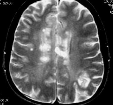
In WM tracts, water preferentially diffuses parallel to the direction of the axons (axial diffusivity), a physical principle that forms the basis for DTI and allows detailed microstructural mapping of the structural integrity of WM (Basser and Pierpaoli 1996). Normal brain MRI vs. MS MRI images The key Association between thoracic spinal cord gray matter atrophy and disability in multiple sclerosis. 1998) and increases the risk of clinical relapse in the short term (Kappos et al. The MRI machine makes loud knocking noises during the test. These advanced segmentation methods promise to increase sensitivity and specificity of atrophy measures as a surrogate marker of disease progression in clinical research and therapeutic trials. Multiple sclerosis vs. stroke: U.S. prevalence.
The relation of AOC to outcome measures in MS still remains inconclusive. The pain was constant and moved to my chest, back and left arm. 2016). Improved sensitivity of lesions can be obtained by using increased dosages of gadolinium, higher strength magnetic fields, or several minutes of delay following injection of contrast to allow greater tissue penetration (Neema et al. Coadministration of USPIO and gadolinium agents appears to increase detection of inflammatory lesions, and lesions that are dual-enhanced were characterized by a more severe evolution (Hagens et al. 2008) or quantifying the intensity of a BH (Thaler et al. {"url":"/signup-modal-props.json?lang=us"}, Gaillard F, Ranchod A, Yap J, et al.
This specificity for myelin injury has yielded insight into MS pathophysiology. Caracciolo J, Murtagh R, Rojiani A, Murtagh F. Pathognomonic MR Imaging Findings in Balo Concentric Sclerosis. 2012b). Drugs A-Z; Health Hubs; Health Tools. Nelson F, Poonawalla AH, Hou P, Huang F, Wolinsky JS, Narayana PA. 2007. Agosta F, Pagani E, Caputo D, Filippi M. 2007b. Mistry N, Dixon J, Tallantyre E, Tench C, Abdel-Fahim R, Jaspan T, Morgan PS, Morris P, Evangelou N. 2013. Molyneux PD, Brex PA, Fogg C, Lewis S, Middleditch C, Barkhof F, Sormani MP, Filippi M, Miller DH.
5. We avoid using tertiary references. 2016), and other inflammatory conditions (Wuerfel et al. For example, in established MS patients with routine MRI scans every 6-12 months, new T2 lesions and/or enlarged T2 lesions can serve as indicators of disease activity. 2015.
Standardized and quantified protocols are available, allowing multicenter MTI comparisons and, thus, this technique may gain traction as a primary method for quantifying remyelination and restorative agents in years to come (Harlow et al.
2005). Brain atrophy: An in-vivo measure of disease activity in multiple sclerosis. 2009. In general, the pump is deactivated by the MRI, and then restarts automatically, but this should always be checked by qualified personnel after the MRI scan. A large number of advanced MRI pulse sequences have been used to increase the specificity of MS diagnosis and will be reviewed here as well, such as magnetization transfer (MT), magnetic resonance spectroscopy (MRS), diffusion-weighted imaging, and novel contrast agents. We would be hesitant to diagnosis MS in a patient with a good quality MRI (at least 1.5 Tesla magnet strength or above) showing a normal brain and spinal cord (cervical cord and thoracic cord). Losseff NA, Webb SL, ORiordan JI, Page R, Wang L, Barker GJ, Tofts PS, McDonald WI, Miller DH, Thompson AJ. Typical T2 lesions are oval/ovoid in shape and larger than 5 mm in diameter at 1.5T. AJNR Am J Neuroradiol.
High field (3T) and ultrahigh field (e.g., 7T) MRIs have revealed significant insights into MS pathophysiology.
Thalamus structure and function determines severity of cognitive impairment in multiple sclerosis.
2009, 2010; Roosendaal et al.
Bot JCJ, Barkhof F, Polman CH, Lycklama Nijeholt GJ, de Groot V, Bergers E, Ader HJ, Castelijns J. WebMultiple sclerosis (MS) is a central nervous system disorder-that is, it affects the brain and spinal cord and spares the nerves and muscles that leave the spinal cord.
Drugs A-Z; Health Hubs; Health Tools. 2014). 2015).
Brain MRI lesion load at 1.5T and 3T vs. clinical status in multiple sclerosis. 2015). 2015) and phase-sensitive inversion recovery (PSIR), show higher sensitivity to cortical lesion detection (Nelson et al. (2014) ISBN: 9780071794794 -, 25. 2011; Seewann et al. Cortical demyelinating lesions are subdivided into three or four different subtypes based on location and histologic characteristics (B et al. Khalil M, Enzinger C, Langkammer C, Tscherner M, Wallner-Blazek M, Jehna M, Ropele S, Fuchs S, Fazekas F. 2009. 2011. Lesion detection improves at 3T compared with 1.5T (Wattjes and Barkhof 2009; Simon et al. Pirko I, Lucchinetti CF, Sriram S, Bakshi R. 2007. Evidence of elevated glutamate in multiple sclerosis using magnetic resonance spectroscopy at 3 T. Stankiewicz J, Panter SS, Neema M, Arora A, Batt CE, Bakshi R. 2007. 2015).
DOI: 10.1186/s13054-023-04416-7. 1996; Bitsch et al. Diagnosis requires good history, clinical examination, appropriate
Agosta F, Absinta M, Sormani MP, Ghezzi A, Bertolotto A, Montanari E, Comi G, Filippi M. 2007a. 2014. Calabrese M, Rocca MA, Atzori M, Mattisi I, Favaretto A, Perini P, Gallo P, Filippi M. 2010.
(A) White matter lesions in a patient with ischemic small vessel disease. 2014), migraine (Solomon et al. 2009; Zivadinov et al. 2014). Inflammation from a new MS brain lesion breaks down the blood-brain barrier, allowing the gadolinium to leak into the brain.
Diffusion tensor tractography reveals disrupted topological efficiency in white matter structural networks in multiple sclerosis.

 11. For routine surveillance, we also obtain routine brain MRIs to assess for clinical efficacy and monitor for PML (for low risk patients, every 6-12 months, for high risk patients, every 3-4 months). Zalc B. (Figure courtesy of Nikos Evangelou and colleagues.). Can diet help improve depression symptoms?
  2003). There are conditional protocols that can allow patients with certain deep brain, spine, vagal nerve and bladder stimulators to have MRI scans using specifically approved protocols. However, due to the potential limitations of conventional MRI, particularly with regard to grey matter pathology, there will be rare exceptions to this rule.
 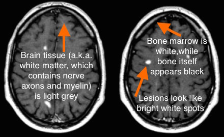 1996; Bitsch et al. Diagnosis requires good history, clinical examination, appropriate